In dentistry, it is necessary to work in confined areas where good visibility is often extremely limited when examining and treating teeth and gums.
For this reason, you have probably noticed many dentists wear loupes, special glasses that provide magnification while working. These are great for many situations, but there are times when greater magnification is needed. This is when we will use a dental microscope.
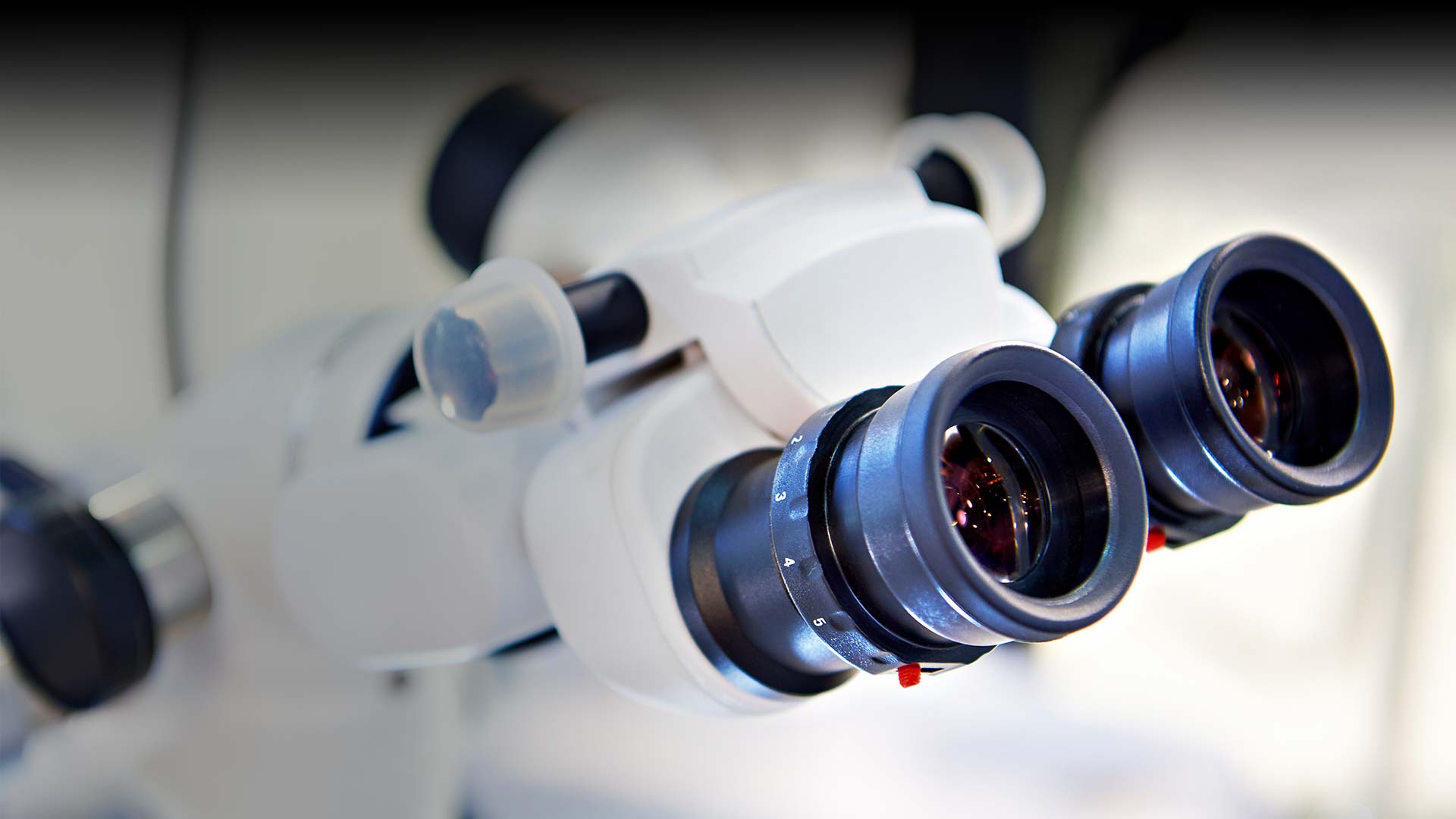
What is a Dental Microscope?
A dental microscope can be mounted onto the wall or on a movable arm, so we can easily adjust it, providing an excellent view of a patient’s mouth. It has a bright LED light, so we can clearly visualize the inside of the mouth without any shadows.
Our dentists can easily change the magnification level and bring even small areas into sharp focus. The microscope has special filters that can be turned on for specific procedures.
What are the Benefits of Microscopic Dentistry?
There are several benefits to using a dental microscope, including the following.
Better Depth of Focus
Dental microscopes have different levels of magnification, so we can zoom in and out to view specific areas of the mouth more clearly, and different dental procedures often require different levels of magnification.
Improved Illumination
Because the mouth is quite dark, it is frequently tricky to angle a light in precisely the right direction. The LED light on a microscope is on the same axis as the area we need to examine, providing an excellent level of illumination.
Faster, More Accurate and Earlier Diagnoses
As you can imagine, improved magnification enables us to view teeth and other structures in much greater detail, which is essential for making quick and accurate diagnoses, often at an earlier stage, as demonstrated recently by Dr. Al Falsafi. Dr. Al examined a patient’s mouth using microscopic dentistry and could see a tiny crack in a tooth that would be impossible to detect otherwise. Dr. Al was then able to restore it before the tooth was damaged more substantially.
A small crack in a tooth is simple to mend with restorative dentistry, and at this stage, treatment is cheaper, quicker and less invasive. Conversely, when a tiny crack goes undetected, it can open slightly every time you bite or chew on that tooth, allowing bacteria to enter the tooth where they can cause infection and decay. That tiny crack could eventually result in a severe tooth infection requiring root canal therapy or, worse, result in tooth loss.
Patient Education
We can take pictures or a video during an exam. These images can be extremely useful for patient education so you can see what we see, allowing us to explain certain problems more easily, so you can understand the issue and recommended treatment. Images can be emailed to a dental specialist if we require a second opinion or need to refer a patient to a dental specialist, speeding up the process.
Improved Precision
Viewing a tooth in greater magnification enables our dentists to prepare and precisely fit dental restorations. We can also finish restorations more easily, potentially improving overall aesthetics.
From our point of view, using microscopic dentistry at Bel Canto enables our dentists to sit more comfortably during dental exams and treatments, helping avoid back issues. However, we love this technology because it allows us to provide our patients with a better, more accurate service. Nothing can give us greater satisfaction than treating a small issue more quickly and effectively and sending our patients home with healthy, attractive smiles.